Guided endodontic access in a calcified tooth


This case was referred to me for a root canal treatment of a lower left central incisor (31).
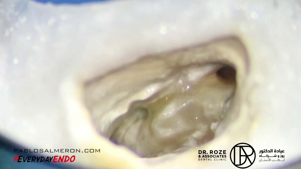
The tooth was showing a chronic apical periodontitis, likely due to a previous trauma. As a result, a calcific metamorphosis, or canal calcification, was present.
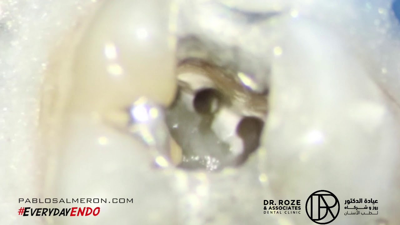
The complexity of the case lay in the calcification of the canal in a very narrow root, making the treatment challenging.
Since half of the canal was calcified, the freehand approach carried significant risks. Even a slight deviation in the trajectory during the access could lead to a perforation. Therefore, a guided endodontic access was planned.
To create the guide, I used information from a CBCT (3D x-ray) and an intraoral scan (3D modeling of the teeth). Using specialized 3D modeling software, a digital model of the mouth was recreated.
With the aid of the guide, the root canal treatment was successfully completed, and the canal calcification was effectively managed.
This highly complex case was successfully resolved thanks to meticulous digital planning and the use of guided technology.
With the right experience, meticulous planning, and the appropriate tools, complex cases can often be successfully managed. I always recommend seeking the expertise of a specialist with experience in handling such situations to prevent further damage and achieve the best possible outcome. While individual results may vary, the success rate is highly predictable in the vast majority of cases.
Let me help you save your tooth.
Plans are of little importance, but planning is essential.
PhD. MSc. Dr. Pablo Salmeron.