90 degrees roots. Root canal treatment.

– Second lower molar with 90 degrees twisted roots.
– Labomed microscope.
– MTwo + Reciproc files.
PhD. MSc. Dr. Pablo Salmeron.
Consultant Endodontist
– Second lower molar with 90 degrees twisted roots.
– Labomed microscope.
– MTwo + Reciproc files.
PhD. MSc. Dr. Pablo Salmeron.
Successful root canal therapy requires an excellent knowledge of both the external and internal anatomy of root and its canal morphology. Extra roots or root canals, if not detected, are a major reason for endodontic failure. Maxillary molars show considerable anatomic variation and abnormalities with respect to the number of roots and root canals. Traditionally, the maxillary second molar has been described to have 3 roots with 3 or 4 root canals, with the fourth canal commonly being found in the mesiobuccal root (MB2). Several authors have reported of maxillary second molars presenting with 4 roots with the accessory root being the second palatal.
In this case, an upper second molar, I found a 4th canal in an unsual location.
A second palatal canal or a centered MB2? Watch it in HD 1080p.
– Second upper molar with an unusual 4th canal.
– Labomed microscope.
– MTwo + Reciproc files.
PhD. MSc. Dr. Pablo Salmeron.
Root canal treatment, first upper molar.

– First upper molar root canal treatment.
– Labomed microscope.
– MTwo + Reciproc files.
PhD. MSc. Dr. Pablo Salmeron.
Root canal treatment. Second molar. Endodontist.

– Lower second molar root canal treatment.
– Labomed microscope.
– MTwo + Reciproc files.
– Sony Alpha 5000.
PhD. MSc. Dr. Pablo Salmeron.

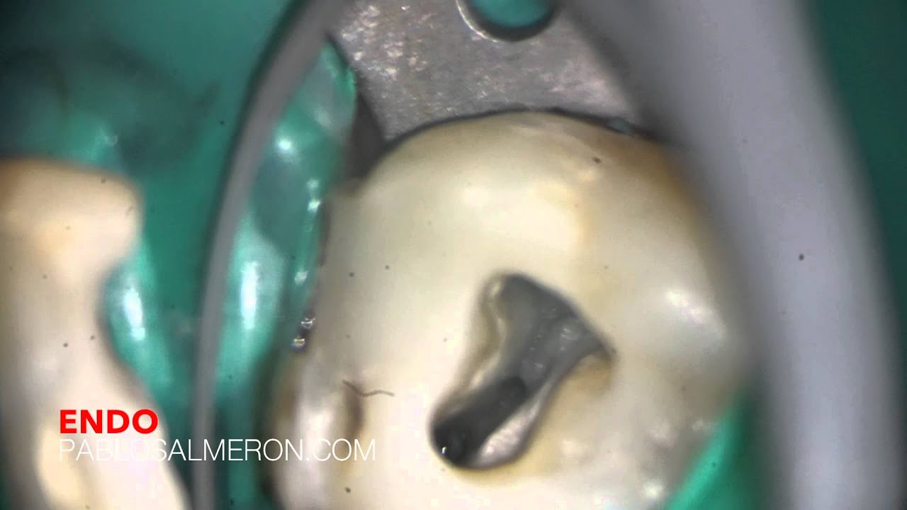
Para los que no sois odontólogos os explico. Lo que veis es la cámara pulpar de una muela. Los agujeros negros son los conductos radiculares, son como pelos de finos, y recorren cada una de las raíces.
En la cámara pulpar se encuentra un paquete vasculonervioso que irriga el diente. Lo que yo hago como endodoncista y con ayuda de un microscopio es una microcirugía en la que extraigo todo ese paquete, compuesto por arterias, venas y tejido nervioso, para luego sellar con un material que inyecto a 300 grados en cada conducto.
– Labomed microscope.
– Sony a5000.
– Mandibular first molar pulp chamber.
Dr. Pablo Salmerón.