Pulp Horn Amputation

Today’s case is a 11 year old patient with a big caries on the second maxillary molar referred to me to save the tooth. My duty as endodontist is not only providing root canal treatments but also preserving teeth from them too.

In this particularly case I performed a pulp horn amputation.

pulpotomy

These are the steps taken:

– Rubber dam isolation.

– Microscope magnification.

– Manual removal of the soft dentin with excavator.

– Pulp horn amputation.

– Disinfected and rinsed with NaOCl from “outside” to achieve haemostasis.

– Dried with sterile cotton pellets.

– Bioceramic capping.

– Direct glass ionomer as temporally filling on top.

Dentistry today is about the small details.

PhD. MSc. Dr. Pablo Salmeron.

Re root canal treatment upper second molar

This is a re-treatment of an upper second molar #27 through an Emax crown . These kinds of treatments are difficult. However, sometimes they can be easy, and other times they can be impossible. The re-treatment of the tooth depends a lot on the work carried out by the previous dentist therefore, I endeavour to be honest with my patients, as sometimes I am not able to fix everything.

In the following case I had to re-treat a tooth filled with Thermafil. Although Thermafil is a great system and works very well in the hands of some endodontist, it doesn’t work for me. I feel that I can’t control the obturation in 3D and the plastic carrier can be easily exposed without guttapercha around.

For that reason, I feel much more comfortable and secure using vertical condensation with System B, it requires maybe more training under the microscope but is worth it and works really well for me.

27reendo

– Tooth #27

– Microscope

– K-Files #10 #15 #20

– Wave One Gold

– Bioceramics + System B.

Never give up, because when you think it’s all over, is the moment where everything starts.” — Jim Morrison.

PhD. MSc. Dr. Pablo Salmeron.

The Palestinian International Dental Conference

title conferenceLast month I gave a lecture at The Palestinian International Dental Conference. I have to start this post thanking particularly to the BAIRD Academy and the Palestine Dentistry Society for inviting me to the congress and make me feel like I was at home. I want to thank especially Dr. Hassan Maghaireh, Dr. Raed Junaidy, Dr. Anan Amro, and Dr. Ahmad Abd El-Ghani for all their help and for showing me the most beautiful areas of Jordan, Palestine and Jerusalem.

The topic I covered was about Magnification, Cumulative Trauma Disorders (CTDs), Ergonomics and 4 Handed Workflow. For me, all those 4 concepts, are intimately related.

poster
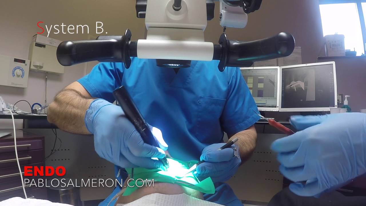
Our manual capacity is limited by our own eyes. It’s because of that why we cannot be more precise, simple as that. To be able to see fine details, we have to bring the object closer to the eye. This is why dentists have to bend down over the patient’s mouth, and this is why dentist have back problems.

The use of magnification is the way to overcome our eye’s limitations. This will also make us work in a neutral and healthy position resulting in long lasting professional career.

4 handed workflow is the masterpiece which completes the ergonomic puzzle. Here you have a couple of videos where I show basic movements which you can start using till you develope your own technique. The first video has been recorded in 30 minutes with an assistant with no previous experience at all; and the second one with a new assistant after one month training. Watch in HD.

YouTube player

YouTube player

“Make it a point to do something every day that you don’t want to do. This is the golden rule for acquiring the habit of doing your duty without pain.”  Mark Twain.

PhD. MSc. Dr. Pablo Salmeron.

Importance of discipline taking records

waxonwaxoffDiscipline is any training intended to produce a specific character or pattern of behaviour. Discipline is nothing but point a goal and achieve it through a protocol, discipline is consistency, discipline is hit the stone always in the same place, always with a good guidance of course.

We live in an age where there aren’t dragons to slay or lands to conquer, and where access to resources and opportunities are so abundant that to know what we want and go after it’s the only gap from heroism. Today the (main) problem isn’t that you can’t get what you want, but you don’t want it enough. Most things we want to achieve aren’t difficult, it’s only lack of desire or fear what separate us from it and it’s at that point where discipline plays a key role.

I have always considered myself a very disciplined person, but discipline doesn’t mean success if isn’t focused on the right direction. A high-level athlete needs self-discipline but also a coach to motivate and guide him through the whole process, no one born knowing everything.

Let’s transfer this to dentistry and the topic I want to address, “the importance of discipline taking records“. A long-term success treatment is based on good planning, today we know that this is not applied in everyday dentistry. We diagnose fast and wrong without a good analysis of the situation; in the same way we live fast our live. This is often because of fear of understanding the complexity of the case or because we’re afraid that the patient won’t accept treatment. If we’re at that point, we are doomed to failure.

We have to understand that being in a situation of discomfort is good, it makes us be more aware, it makes us want to investigate and understand what’s out of our control. To be in a discomfort situation makes us progress!

To understand what’s happening in the mouth is essential to design a good treatment plan, it’s critical for long term success, it’s essential to avoid unexpected surprises and is essential to the patient. The first step in all this starts with taking good records, we must be careful, we must be precise, there are too many steps in the process in which we can miss some information, so the closer we are to perfection, the better will be our job.

Understand all this has taken me a while, and it was not until I started working with Dr. Ian Buckle when I started to fit the pieces of the puzzle, “discipline is perfection Pablo, we must be precise!“.

We work in a multidisciplinary team in which excellent records is critical. What for me may not be relevant as endodontist can be critical for the orthodontist or the surgeon, details are perfection.

We can have many years of experience, we can have a lot of knowledge in our field, but if we don’t understand that a good treatment plan begins with good records, we are doomed to be an average dentist.

Discipline is the bridge between goals and accomplishment“. Jim Rohn.

PhD. MSc. Dr. Pablo Salmeron.

The importance of an accessory screen in endodontics

20150924_161013_resized In this post I would like to talk about why an accessory screen, as much bigger the better, is very important for me on a root canal treatment.

Documentation: An accessory screen allows you to take better photos and videos. One disadvantage of microscopes that do not have built in cameras and need an accessory camera is the difficulty of “pairing” what you see through the eyepieces and what your camera captures through the beam splitter. That image you want to capture and document, should be the same focused image through your eyes and through the camera, but requires a perfect configuration thereof, and the correct setting of the diopter correction of the eyepieces. The camera captures the image of one eye, either the right or left depending in which side of the microscope the camera is. When we look through the microscope we have stereoscopic vision, we see with both eyes at the same time, and is our brain with his own corrections who made a sharp image in the end. The problem here is that without that perfect “pairing” between camera, eyepieces, eyes and brain if we need to document the most tiny detail, that is only focused in one particular millimeter depth of field, we may find that the image of the detail that we wanted to document is not focused at all.

One of the options is trying to focus through the LCD of the camera, the problem is that the size of the LCD is small and usually is in a position that is not comfortable to focus through it.
For me the solution was to connect the microscope to a 55″ screen that I have in front of me. Through that screen it is very easy to document every detail and get good quality photos and videos with the details that I want to show totally focused.

Assistants: Working with a large screen allows my assistant see what I see so she can help me better. Your assistants are part of the treatment, involve them and show what you see motivates them to work. Another advantage is that they can avoid look directly to the work field for more time. This prevents from suffering headaches and vision problems as the light source of the microscope is very bright and brings all these problems after a long work session.

Education: A large screen allow me to teach and train other dentists who want to learn about endodontics.

These are the three reasons why I love working with a large accessory screen.

PhD. MSc. Dr. Pablo Salmeron.