Blog_Pablo_Cover_1
Image is not available

Blog and Cases

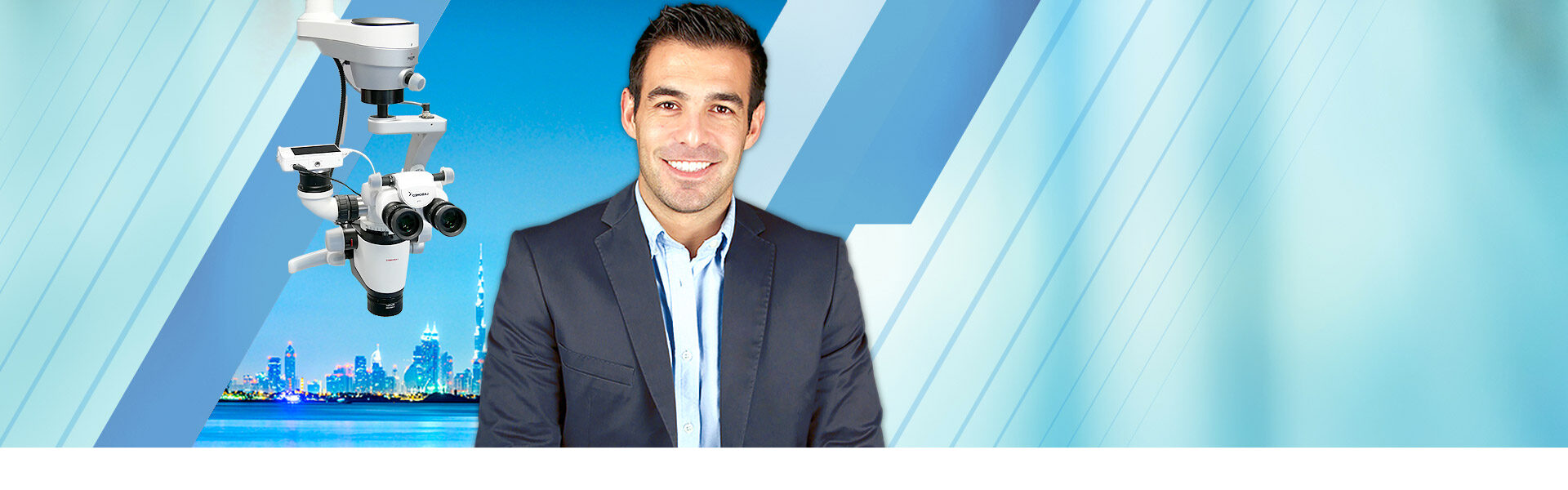
Microscope hands on course in your own practice

brochure hands on
I would like to introduce you this course I teach about the use of the microscope in your own practice. This course is recommended for those dentist who own a microscope (the brand doesn’t matter) and want to improve their technical skills with it.

The course takes place in your own practice with your own team and assistants, in your own space and with your own patients, so you will be able to maximize the use of the microscope making a pleasant working experience.

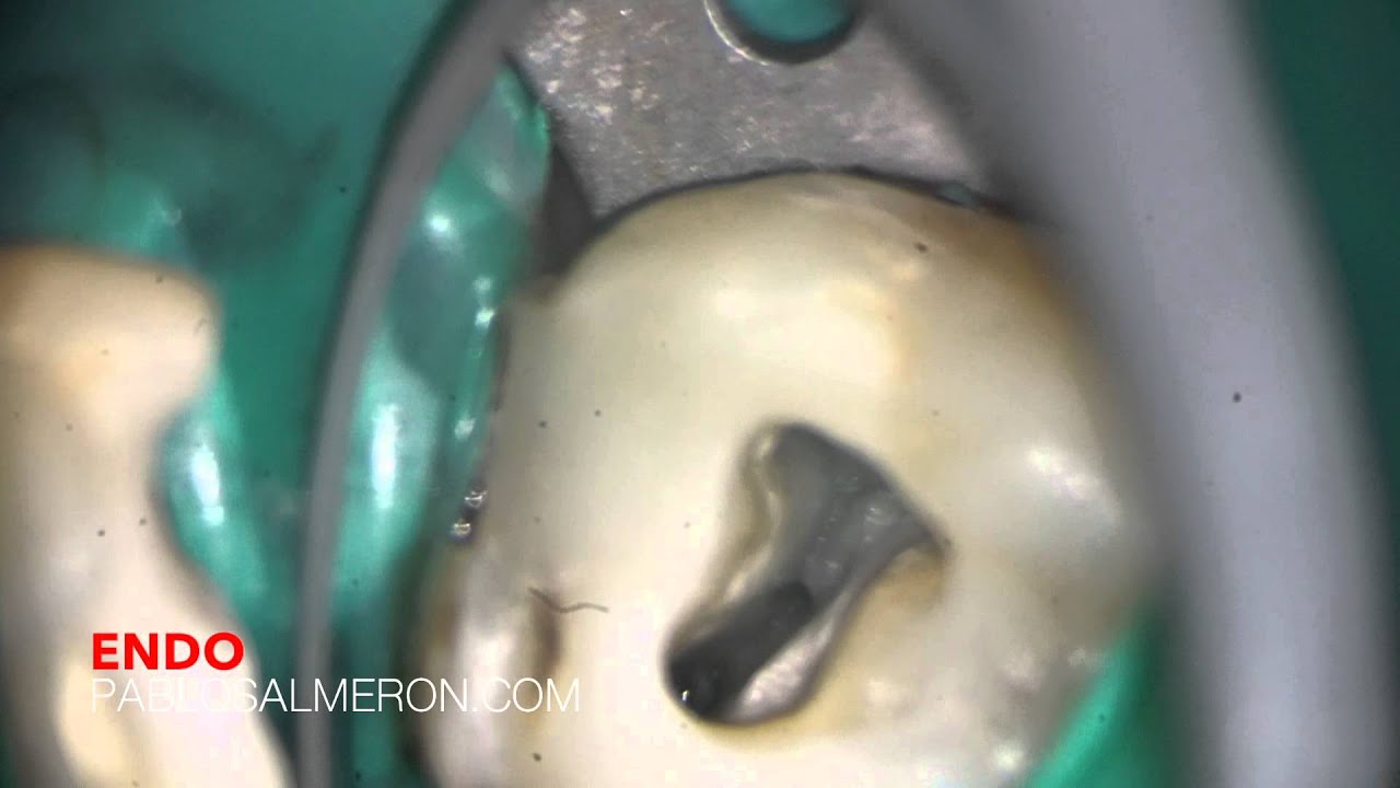
You can only treat what you see.

PhD. MSc. Dr. Pablo Salmeron.

A second palatal canal or a centred MB2?

Successful root canal therapy requires an excellent knowledge of both the external and internal anatomy of root and its canal morphology. Extra roots or root canals, if not detected, are a major reason for endodontic failure. Maxillary molars show considerable anatomic variation and abnormalities with respect to the number of roots and root canals. Traditionally, the maxillary second molar has been described to have 3 roots with 3 or 4 root canals, with the fourth canal commonly being found in the mesiobuccal root (MB2). Several authors have reported of maxillary second molars presenting with 4 roots with the accessory root being the second palatal.

In this case, an upper second molar, I found a 4th canal in an unsual location.

A second palatal canal or a centered MB2? Watch it in HD 1080p.

YouTube player

– Second upper molar with an unusual 4th canal.

– Labomed microscope.

– MTwo + Reciproc files.

PhD. MSc. Dr. Pablo Salmeron.