Blog_Pablo_Cover_1
Image is not available

Broken file retrieval on an upper second molar

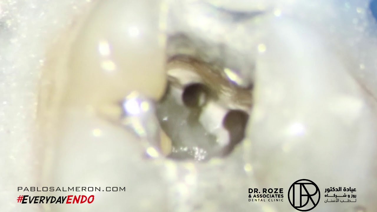
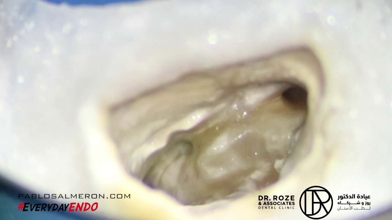
This patient was referred to me for a re-root canal treatment of an upper left second molar (27). This tooth presented a broken instrument in the palatal canal. Due to the location of the tooth (the last one) and the location of the broken file at the apex, the treatment became an important challenge. For these types of cases, I always take a CBCT to properly plan the treatment.

value honesty with all my patients, and I always explain the challenges of each case and the possible outcomes to them. With the right experience, planning, and tools, complex cases are typically successful. My advice is always to seek out an experienced specialist to address these situations and prevent further damage.

While results may vary from patient to patient, they are highly predictable in the vast majority of cases.

Keep challenging yourself to think better, do better, and be better.

PhD. MSc. Dr. Pablo Salmeron at Dr. Roze & Associates.

Removal of silver points

This patient was referred to me for a re-root canal treatment of an upper left second premolar (15). The tooth was presenting a couple of metal post and a silver point obturation. Within the always difficulty of  re-root canal treatments, removal of metal posts and silver points are quite predictable with the proper skills. This tooth was already treated 2 times in the past so preserve as much natural tooth structure was very important. Every time that the tooth is root canal treated or re-treated adds another layer of difficulty for the next treatment making very challenging sometimes to fix what it was done 20 or 30 years ago. My advice is always to find the right specialist with experience to fix this kind of situations to avoid further damage. 

The obturation was done using bioceramic sealer and the treatment was completed in one session. The result may vary from patient to patient but it’s very predictable in the vast majority of cases. Let me save your tooth

Patience and perseverance have a magical effect before which difficulties disappear and obstacles vanish.

PhD. MSc. Dr. Pablo Salmeron at Dr. Roze & Associates.

Upper first molar with 5 canals, MB3

This is why the use of the microscope is so IMPORTANT in Endodontics. This is an upper first molar with 5 canals. I like to clean and shape first the main canals to work after on the accessories. In this case I found a MB2 and MB3 canal. It’s IMPOSSIBLE to do this with the limitations of our naked eye, even with loupes. Mind as well the “conservative” access done in this case.

YouTube player

Love looks through a telescope; envy, through a microscope.

PhD. MSc. Dr. Pablo Salmeron at Dr. Roze & Associates.

The power of magnification

I can’t imagine to do a root canal treatment without my microscope. The human eye is limited and without microscope, we are not able to see important details for the long term success of the treatment.

This photo shows different grades of magnification. It was taken before the obturation with the canals already cleaned and shaped that’s why the MB2 is so visible.

Vision is the art of seeing what is invisible to others.

PhD. MSc. Dr. Pablo Salmeron at Dr. Roze & Associates.